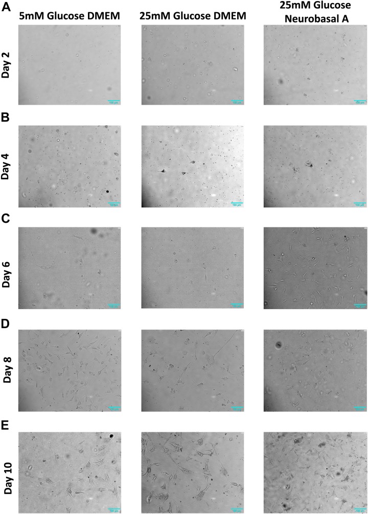
FIGURE 1.
Morphology of retinal neurons cultured in 5 mM glucose DMEM, 25 mM glucose DMEM, or 25 mM glucose Neurobasal A medium. (A) On Day 2, adherent cells are few in number in all three media conditions. (B) On Day 4, more cells start to attach to the cell culture dish in 25 mM glucose- containing DMEM and Neurobasal A medium, whereas fewer cells are adherent in 5 mM glucose containing DMEM. (C) On Day 6, more cells start to adhere to the culture dish, especially in high glucose-containing Neurobasal A medium. (D) Day 8 adherent cells continue to grow in culture. (E) On Day 10 post primary culture initiation, axon-like projections were prominent in length, gradually forming complex networks. N = 5.

