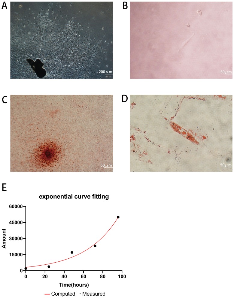
Figure 1.
Isolation and characterization of GMSCs. (A) Cultured primary GMSCs from gingival tissues have a long spindle shape. Scale bar: 200 μm. (B) Single primary cells. Scale bar: 50 μm. (C) The formation of mineralized nodules. Scale bar: 50 μm. (D) Accumulated lipid droplets. Scale bar: 50 μm. (E) PDT of GMSCs.
Abbreviation: GMSCs, gingival mesenchymal stem cells.

