Figure 3.
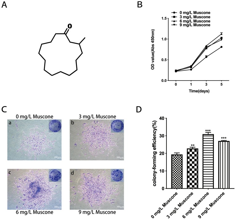
Effect of muscone on proliferation. (A) The chemical structure of muscone (3-methylcyclopentadecanone). (B) GMSCs were treated with 0, 3, 6, and 9 mg/L muscone for 0, 1, 3, and 5 days, and the number of viable GMSCs was analyzed using a CCK-8 assay. Data are presented as the mean ± S.E.M. ***P<0.001 indicates a significant difference between groups. (C) After 10 days, the cells were stained, and more and larger cell colonies were observed in the experimental groups (b, c, d) than in the control group (a). Scale bar: 200 μm. (D) Colony formation assays showed a significant difference between the control group (0 mg/L) and the experimental groups (3, 6, and 9 mg/L muscone). ***P<0.001, **P<0.01.
Abbreviations: GMSCs, gingival mesenchymal stem cells; CCK-8, cell counting kit-8; S.E.M., standard error of the mean.

