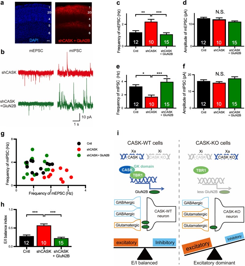
Fig. 5.
Co-transfection with GluN2B rescues the disrupted E/I balance in the spontaneous synaptic transmission in CASK-KD neurons. a Histological images of shCASK + GluN2B-transfected somatosensory cortex. The laminar structure was visualized by DAPI staining (left). tdTomato-labeled shCASK + GluN2B-transfected neurons were in layer 2/3 of the somatosensory cortex (right). Cortical layers are indicated by numbers. Scale bar: 100 µm. b Representative traces of the mEPSC (left) and mIPSC (right) from shCASK neurons (top) and shCASK + GluN2B neurons (bottom). Scale bars represent 10 pA (vertical axis) and 1 s (horizontal axis). c, d Frequency (c) and amplitude (d) of the mEPSC in control (black), shCASK (red), and shCASK + GluN2B (green) neurons. GluN2B transfection rescued the increased mEPSC frequency in shCASK-transfected neurons (animal numbers; Cntl n = 3, shCASK n = 3, shCASK + GluN2B n = 3). e, f Frequency (e) and amplitude (f) of the mIPSCs in control, shCASK, and shCASK + GluN2B neurons. GluN2B transfection rescued the decreased mIPSC frequency in the shCASK-transfected neurons. g. Distribution of the frequency of mEPSCs versus mIPSCs shown in a scatter plot. Each dot represents a single cell. h E/I balance index of neurons with each KD. i Molecular mechanism underlying the disruption of the E/I synaptic balance in CASK heterozygous female mice. Either the CASK-KO or WT allele is randomly inhibited (Xi) by XCI, and the activated X chromosome (Xa) determines the genotype of the cell. In CASK-KO neurons, GluN2B expression is down-regulated, resulting in a shifting of the E/I synaptic balance toward excitatory dominance. Statistical significance was determined by ANOVA and Bonferroni’s post hoc test (c–f and h). *p < 0.05, **p < 0.01, ***p < 0.001. Numbers on bars are the number of cells analyzed. N.S. not significant

