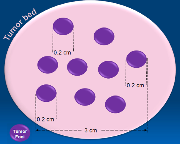
Figure 1.

Conceptual figure showing the dilemma of measuring tumor size, with residual islands of tumor (purple) in a fibrotic tumor bed (beige). In this example, the tumor bed measures larger than the farthest extent between two tumor foci (3.0 cm). Currently, there are no guidelines of how to address the dilemma of measuring tumor size. Many pathologists continue to use gross size (4.0 cm in this example), while others use a mapped grossing approach, which in this case would yield a tumor size of 3.0 cm. In case of a single focus of residual tumor, tumor size is measured microscopically (0.2 cm). Some pathologists like to add up the microscopic dimensions of individual tumor foci (e.g. 0.2 cm + 0.2 cm + …) for the tumor size.
