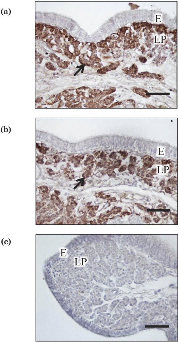
Fig. 3.

Immunostaining for CabP-D28k in uterine tissues incubated with (a) or without (b) IL-6 (100 ng/mL) for 1.5 h. Arrows indicate the presence of immunoreaction products in tubular gland cells. Negative-control staining (using normal mouse IgG instead of primary antibody) did not yield a positive reaction (c). E = mucosal epithelium, LP = lamina propria. Scale bars = 50 µm.
