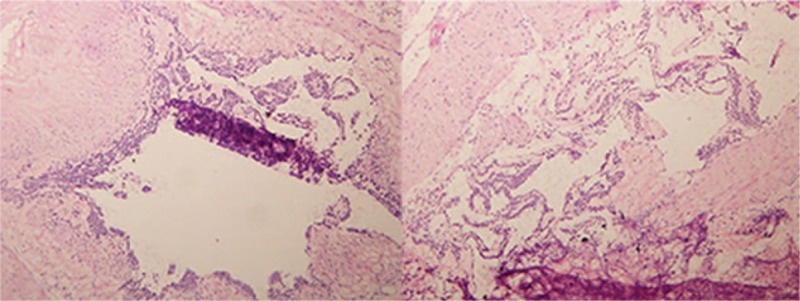
Figure 3.

A partial resection of the distal stomach was examined, showing a mobile, spherical mass of approximately 2× 2 cm, from the medial to the serosal layer in the gastric antrum. No swollen lymph nodes were observed around the stomach. The frozen specimens showed that the tumor was confined in the mucosa with a greater curvature in the gastric antrum (hematoxylin and eosin staining; 100× magnification).
