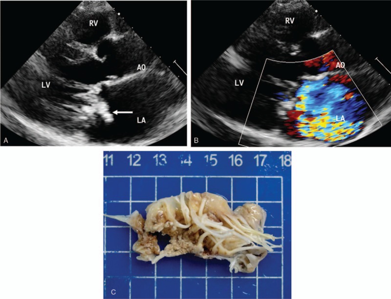
Figure 1.

IE in a 50-year-old man. (A) Echocardiography in the left ventricle parasternal long-axis view shows a large complex vegetation on the mitral valve (arrows). (B) Color Doppler shows severe mitral regurgitation. (C) Specimen shows a large number of vegetations on the mitral valve. AO = aortic, LA = left atrium, LV = left ventricle, RV = right ventricle.
