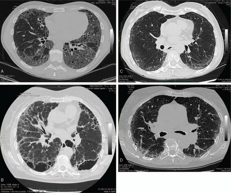
Figure 1.

Representative high-resolution computed tomography images from the subjects with rheumatoid arthritis-interstitial lung disease. (A) A 64-year-old female with fibrotic changes of usual interstitial pneumonia pattern, reticular and ground glass opacity, septal thickening diffusely, and traction bronchiectasis. (B) A 69-year-old male revealing septal thickening diffusely and extensive macrocystic honeycombing. (C) A 65-year-old male with bilateral peripheral ground glass opacity and typical subpleural sparing representing the nonspecific interstitial pneumonia pattern. (D) A 70-year-old female showing evidence of bilateral peripheral reticular and ground glass opacity of the left lower lobe with peripheral consolidations typical of organizing pneumonia.
