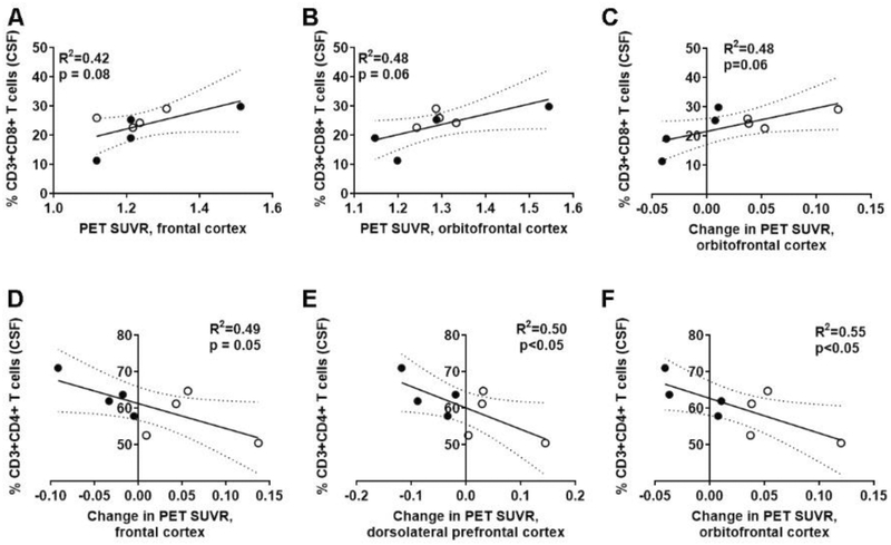
Figure 6. Higher levels of CSF-localized CD8 T cells, and lower CD4 T cells associate with increased Aβ deposition after physical activity.
(A-C) Standardized uptake value ratio (SUVR) for PET imaging of 18F-florbetapir (amyloid burden) associated higher CD8+ T cells in the cerebrospinal fluid (CSF) in both (A) frontal and (B) orbitofrontal cortices after PA. Individuals in the ST (closed circles; n=4) and AET (open circles; n=4) interventions are identified. (C) Increased CD8 T cells also associated with increased Aβ deposition. (D-F) There were concomitant decreases for CD4 T cells in (D) frontal, (E) dorsolateral prefrontal, and (F) orbitofrontal cortices with increased Aβ deposition when comparing baseline and post-PA PET imaging.

