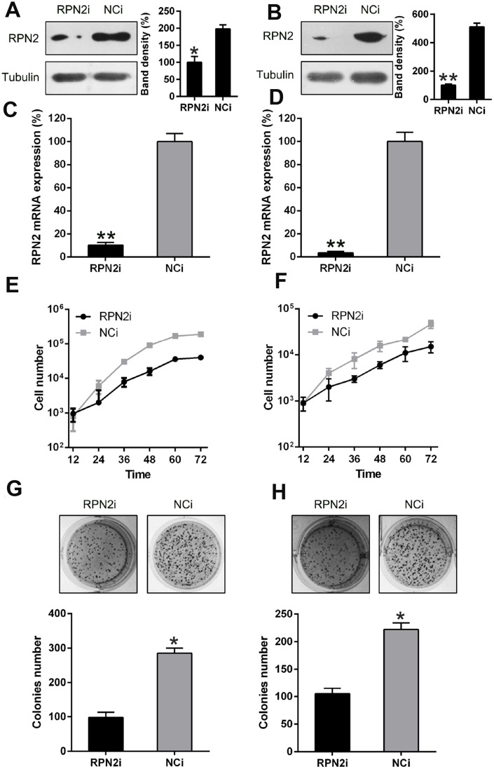
Figure 4.
RPN2 silencing restrained HCC cell proliferation. Western blotting (A, B) and qPCR (C, D) were conducted to confirm the silencing of RPN2 in both cell lines. (E, F) multiplication rate of Huh-7 and HepG2 cells was measured at the time points of 12, 24, 36, 48, 60, and 72 h after the transfection by using MTT assay. (G, H) Soft agar colony formation test was used to examine the replicative rate of HepG2 and Huh-7 cells transfected with shRNA-RPN2. The band of target protein was normalized to the density of action. The quantification was performed independently in a single band. Data are recorded as mean ± SD. *P < 0.05, **P < 0.01, ***P < 0.001 vs control group.

