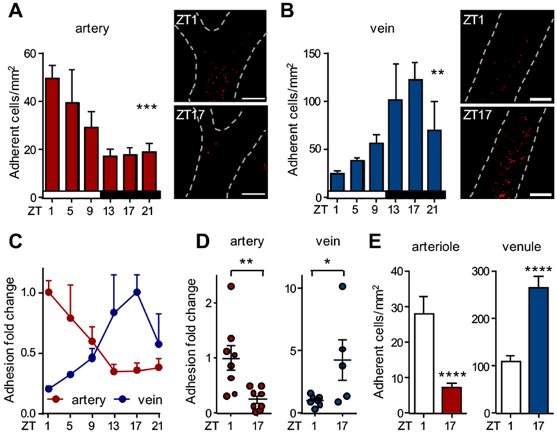
Figure 1. Inflammatory leukocyte adhesion to arteries and veins occurs at different times of the day.

(A-B) In vivo quantification of adherent leukocytes after TNF-α stimulation over 24 h in carotid artery (A) and jugular vein (B); n = 4-13 mice, one-way ANOVA. (C) Normalization of the leukocyte adhesion data from (A-B). Data are normalized to peak levels; n = 4-13 mice. (D) In vivo quantification of adherent cells in Lyz2-gfp mice after TNF-α stimulation in carotid artery and jugular vein. Data are normalized to ZT1 levels; n = 5-8 mice, Student’s t-test (artery) and Mann-Whitney test (vein). (E) In vivo quantification of adherent cells in arterioles and venules of the cremasteric microvasculature after TNF-α stimulation; n = 5 mice, Student’s t-test. *p < 0.05, **p < 0.01, ***p < 0.001, ****p < 0.0001. Scale bars: 200 μm.
