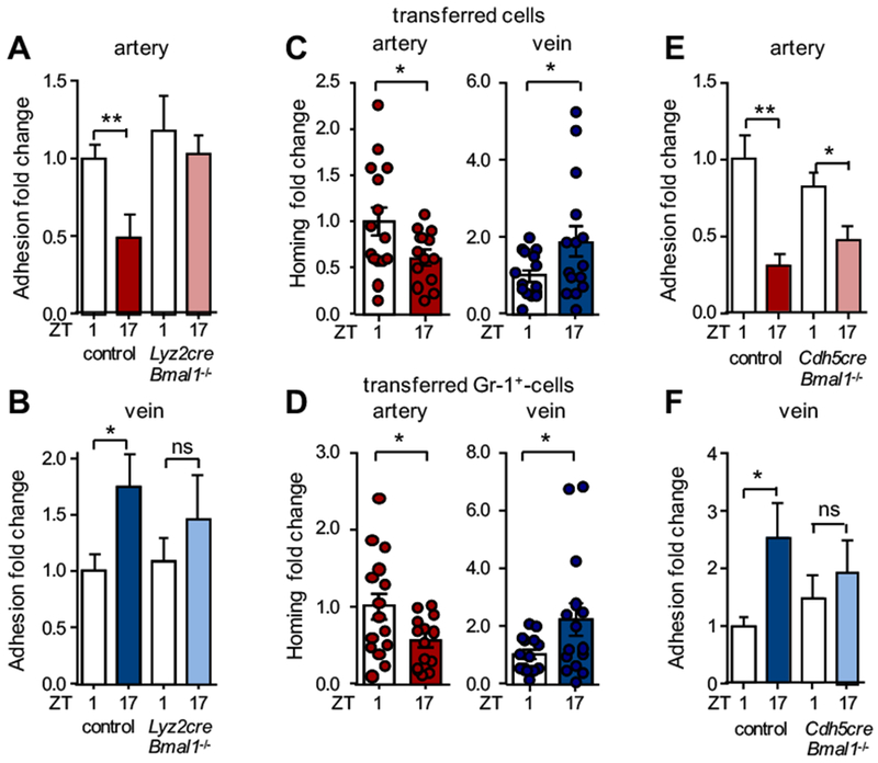
Figure 2. Role of rhythmicity in myeloid cells and the microenvironment.

(A-B) In vivo quantification of adherent leukocytes after TNF-α stimulation in carotid artery (A) and jugular vein (B) in control and Lyz2cre:Bmal1−/− mice. Data are normalized to control ZT1 levels; n = 7-10 mice, Student’s t-test. (C) Homing experiments of adoptively transferred leukocytes (C) and Gr1+ cells (D) to carotid artery and jugular vein after TNF-α stimulation. Data are normalized to ZT1 levels; n = 13-15 mice, Student’s t-test with Welch’s correction. (E-F) In vivo quantification of adherent leukocytes after TNF-α stimulation in carotid artery (E) and jugular vein (F) in control and Cdh5creERT2:Bmal1−/− mice. Data are normalized to control ZT1 levels; n = 8-11 mice, Student’s t-test with Welch’s correction. *p < 0.05, **p < 0.01.
