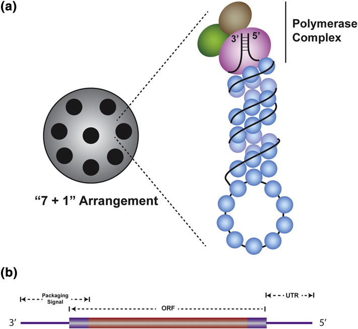
Figure 1. Structure of IAV vRNPs and vRNAs.
(A) Diagram showing the”7 + 1”arrangement of vRNPs inside a virion (left side). A schematic representation of a vRNP complex is shown with a tripartite viral polymerase complex at the top of the vRNP where it binds the panhandle structure formed by 5’ and 3’ UTRs (right side). Rest of the vRNA (black) is covered with multiple nucleoproteins (blue). (B) Schematic diagram showing the positions of packaging signals on a vRNA. The viral ORF is depicted as a rectangle that is flanked by UTRs, which are shown as lines. Typical packaging signals (violet) are situated at both ends of the vRNA consisting of UTRs and ends of the ORF.

