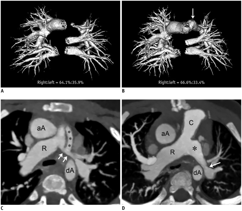
Fig. 3. 1-year-old girl with pulmonary atresia and ventricular septal defect underwent placement of right ventricle-pulmonary artery conduit and left modified Blalock-Taussig shunt before initial cardiothoracic CT.
She underwent unsuccessful surgical left pulmonary artery angioplasty between two serial cardiothoracic CT examinations (group 1B).
A. Frontal volume-rendered CT image of pulmonary vasculature obtained from initial cardiothoracic CT shows segmented right and left pulmonary vascular volumes, and left pulmonary vascular volume is slightly smaller than right side (right:left = 64.1%:35.9%). B. Frontal volume-rendered CT image of pulmonary vasculature acquired from follow-up cardiothoracic CT after surgical left pulmonary artery angioplasty shows unchanged pattern of asymmetrically reduced left pulmonary vascularity (right:left = 66.6%:33.4%), and left pulmonary vascular volume percentage slightly decreased by approximately 2.5%, indicating ineffective surgical left pulmonary artery angioplasty. Even though branch left pulmonary artery is dilated (arrow) as result of surgical procedure, this enlargement does not have effect on CT pulmonary vascular volume percentage. C. Oblique axial CT image obtained before surgical left pulmonary artery angioplasty reveals diffuse narrowing (arrows; 6.7 mm/m2 of body surface area) of branch left pulmonary artery. Left modified Blalock-Taussig shunt (asterisks) is noted. D. Oblique axial CT image acquired after surgical left pulmonary artery angioplasty shows dilated proximal left branch pulmonary artery (asterisk). However, distal segment still shows focal narrowing (arrow; 5.6 mm/m2 of body surface area, approximately 18.5% decrement). C = right ventricle-pulmonary artery conduit, R = right pulmonary artery

