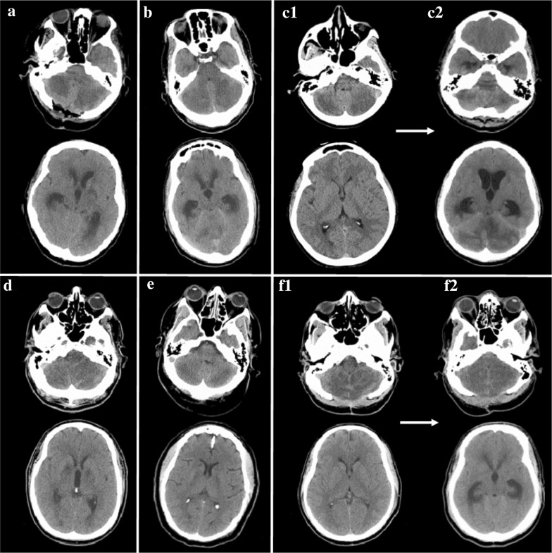
Fig. 1.
Non-contrast computed tomography of the head from patients at the time of arrival or transfer to tertiary care center, at the level of the cerebellum (above) and the lateral ventricles (below). A Patient #1, CT on arrival to tertiary care center, 3 days after initial event; emergent suboccipital decompression was completed on arrival; B Patient #2, at time of presentation, ~ 48 h after last well; C1 Patient #3, on arrival; C2 Patient #3 on hospital day 6, demonstrating evolution of cerebellar edema with obstructive hydrocephalus; D Patient #4, at time of presentation, ~ 24 h from last well; E Patient #5, at time of presentation, ~ 24 h from last well; F1 Patient #4, at time of presentation, ~ 48 h from last well; suboccipital decompression is from a previous event; F2 Patient #4 on hospital day 4, demonstrating evolution of cerebellar edema with obstructive hydrocephalus

