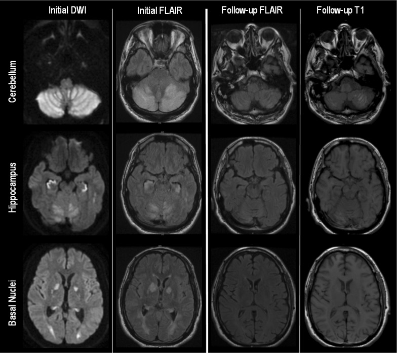
Fig. 3.
Patient #5 MRI brain, selected sequences, initial and three-month follow-up. DWI diffusion-weighted imaging, FLAIR fluid-attenuation inversion recovery, MRI magnetic resonance imaging. Evolution of imaging changes between initial presentation and repeat imaging 3 months later. Abnormal restricted diffusion was not present on follow-up imaging (not shown)

