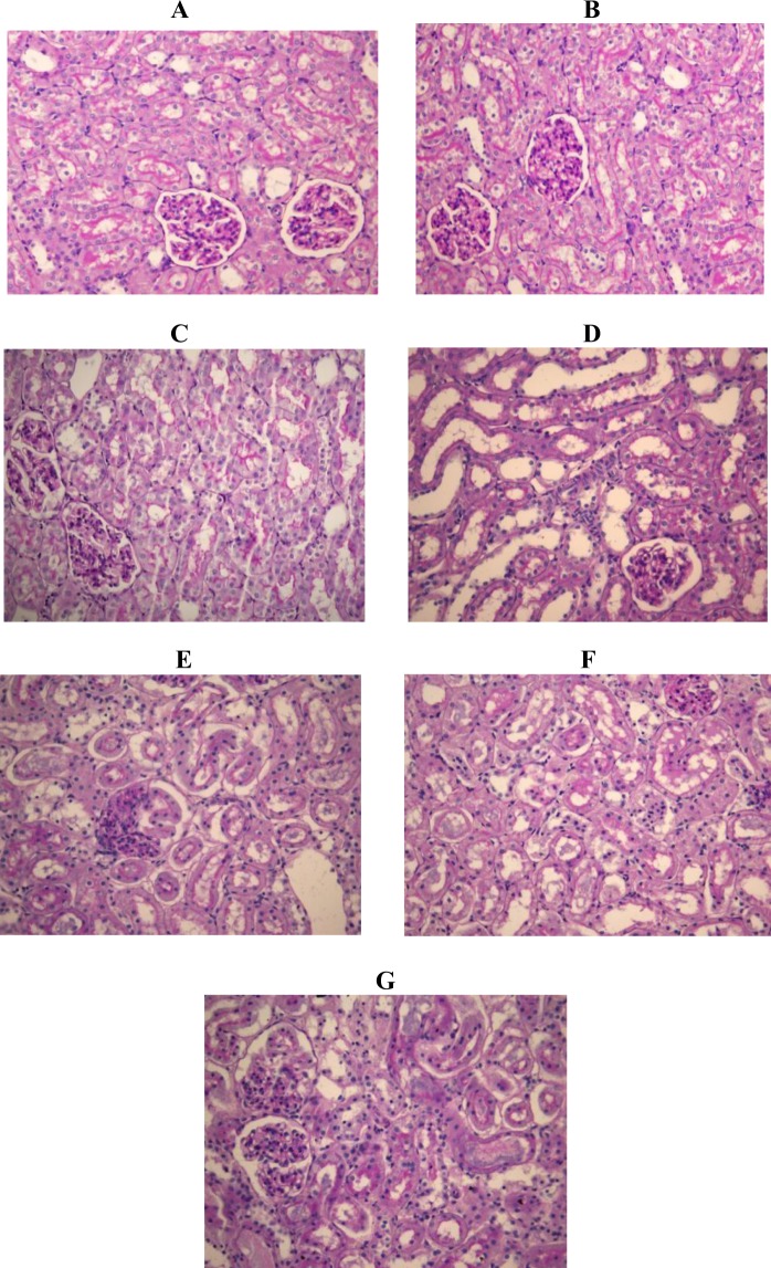
Figure 4.
The effects of pioglitazone (0.3 mg/kg; 1 mg/kg; 3 mg/kg) on histological micrographs of renal tissues. Periodic acid–Schiff (PAS) stain coloring. Original magnification ×20. Figures were randomly chosen from the series of at least 6 experiments (Panels A–G). Panel A: Healthy animal - normal renal parenchyma (PAS staining). Panel B: Animals treated with DMSO only - normal renal parenchyma (PAS staining). Panel C: Animals treated with saline only - normal renal parenchyma (PAS staining). Panel D: Rats subjected to gentamicin-induced nephrotoxicity- marked kidney damage, interstitial edema diffusely present, proximal tubules show loss of brush border and lumen dilatation and loss of nuclei in some epithelial cells. Panel E: Rats subjected to gentamicin induced nephrotoxicity, treated with pioglitazone at dose of 0.3 mg/kg - moderate kidney damage, loss of brush border was observed in half of proximal tubules, in addition to dilatation of lumen and loss of nuclei in some epithelial cells. Panel F: Rats subjected to gentamicin induced nephrotoxicity, treated with pioglitazone at dose of 1 mg/kg –minimal to moderate kidney damage. Panel G: Rats subjected to gentamicin induced nephrotoxicity, treated with pioglitazone at 3 mg/kg – moderate to marked kidney damage, two thirds of proximal tubules show loss of brush border, dilatation of lumen and loss of nuclei in majority of epithelial cells (marked necrosis).

