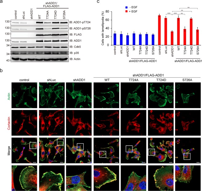
Figure 5.
Phosphorylation of ADD1 at T724 is important for lamellipodia formation. (a) MDA-MB-231 cells were infected with lentiviruses expressing shRNAs specific to ADD1 (sh-ADD1) or luciferase (shLuc). FLAG-ADD1 or its mutants were re-expressed in the ADD1-depleted cells (sh-ADD1/FLAG-ADD1). The cells were serum-starved for 18 h and treated with 40 ng/ml EGF for 15 min. An equal amount of whole cell lysates was analyzed by immunoblotting with the indicated antibodies. (b) The cells as described in (a) were plated on collagen-coated coverslips, serum-starved, and treated with (+) or without (−) 200 ng/ml EGF for 6 h. The cells were fixed, stained for cortactin and F-actin, and visualized with a Zeiss ApoTome2 system. Representative micrographs are shown. Bars, 20 μm. (c) The percentage of cells with lamellipodia in the total number of counted cells was measured (n ≥ 200). Values (means ± s.d.) are from three independent experiments. **P < 0.01; ***P < 0.001.

