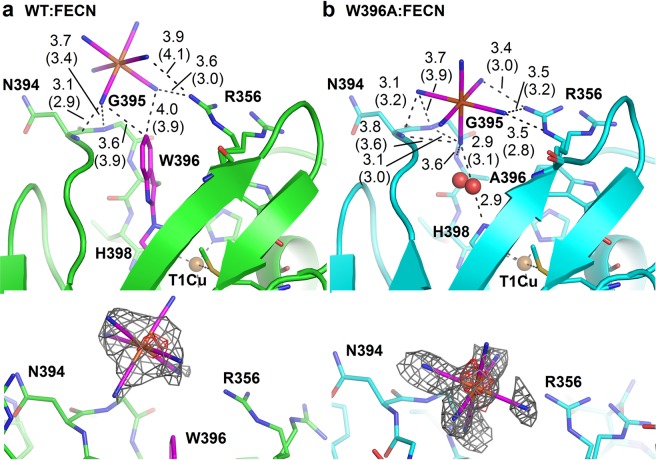
Figure 3.
Binding of ferricyanide in the active site of MvBOx wild type and its W396A mutant. (a) Binding of ferricyanide in OS1 of the WT:FECN structure (6I3J, carbon green). The ferricyanide ion and Trp–His adduct are shown with carbon colored magenta. (b) Binding of ferricyanide (magenta) in OS1 of the W396A:FECN structure (6I3K, carbon light blue). One water molecule (shown as red sphere, in two alternative positions) connects ferricyanide and His398. Interacting residues are marked. Distances are given in Ångströms. If values differ in chain A and B, they are given in parentheses for chain B of the corresponding structure. T1Cu is shown as orange sphere. The composite omit electron density map (2mFo-DFc) is shown as grey mesh and contoured at 1.0 σ level around the ferricyanide ion at the bottom of each panel. The map was calculated using Phenix67. Anomalous difference Fourier is shown as red mesh and contoured at 2.5 σ level around iron. Molecular graphics were created using PyMOL (Schrödinger, LLC).

