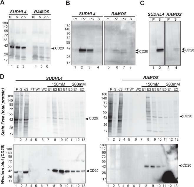
Figure 1.
Expression and CD20 enrichment from SUDHL4 and RAMOS cells. (A) CD20 expression. Cells were harvested from 5 ml of culture medium at 1000 g for 5 min and lysed in PBS + laemmli buffer (1X final). Proteins from cell lysates (10 µl, 5 µl and 2,5 µl, corresponding to 3, 1.5 and 0.75 105 cells respectively) were separated on a 4–15% Tris-glycine SDS-PAGE, transferred to PVDF membrane and immunodetected with an anti-CD20 (1:400) primary antibody and an anti-IgG horseradish peroxidase conjugated secondary antibody (3:10000). (B) Membrane fractionation of SUDHL4 and RAMOS cells expressing CD20. Fractions generated by membrane fractionation of SUDHL4 and RAMOS cells were loaded in a 4–15% Tris Glycine gel (10 µg of total protein for each fraction) and proteins separated by SDS-PAGE (4–15%). The western blot was probed with an anti-CD20 primary antibody (1:200) followed by an anti-IgG horseradish peroxidase conjugate secondary antibody (3:10000). P1, pellet after centrifugation at 1000 g; P2, pellet after centrifugation at 15000 g; P3, pellet after centrifugation at 100000 g (plasma membrane) S, supernatant after centrifugation at 100000 g. (C) Extraction of CD20 from both SUDHL4 and RAMOS cells. Plasma membranes from SUDHL4 and RAMOS cells were solubilized with 1% FC12. After extraction, soluble and insoluble fractions were separated by centrifugation. Proteins from each fraction were separated by SDS-PAGE. The western blot was probed with an anti-CD20 primary antibody (1:200) followed by an anti-IgG horseradish peroxidase conjugate secondary antibody (3:10000). P, pellet; S, supernatant. (D) Enrichment of CD20 using ion exchange chromatography DEAE after solubilization of SUDHL4 and RAMOS plasma membranes. Proteins from pellets (P), supernatants (S), diluted supernatant (dS) flow through (FT), wash (W), elution 1 to 5 (E1 to E5 using 150 mM NaCl) and then elution 1 and 2 (E1 and E2 using 200 mM NaCl) were separated on a 4–15% acrylamide SDS-PAGE. Total proteins were detected after stain-free activation (BioRad method). The presence of CD20 in each fraction was immunodetected with a mouse anti-CD20 (Abcam, 1:200) as a primary antibody and an anti-mouse IgG HRP conjugate as a secondary antibody (3:10000). Full-length gels and blots are included in a Supplementary Information file (Fig. S1).

