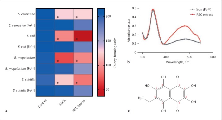
Fig. 6.
Antimicrobial activities of red spherule cell lysates from P. lividus. a Gram-positive (B. megaterium and B. subtilis) and Gram-negative (E. coli) bacteria as well as yeast (S. cerevisiae) were incubated in the presence/absence of the iron chelator EDTA or red spherule cell lysates (2.5 × 10⁵) for 1 h at room temperature. Following treatment, microbes were serially diluted in PBS (pH 7.4) so that 200 colony-forming units were plated onto standard agar medium (LB for bacteria and YEPD for yeast) or agar that had been enriched with iron (Fe3+, ferric ammonium citrate). The colour scale on the right indicates the mean number of viable microbes present on agar, i.e., CFU (n = 3). An asterisk indicates a significant difference (p < 0.05) between the control and treatment groups. b Absorbance spectrum of RSC lysate (∼27 μM echinochrome A) in the absence and presence of 200 μM ferric ammonium citrate. The concentration of echinochrome A in 1 × 10⁵ RSC was calculated by following the extraction method developed by Service and Wardlaw [16] and taking into account that there was ∼6.3 × 10⁵ RSC per mL of P. lividus coelomic fluid. c Molecular structure of the 1,4-napthoquinone pigment, echinochrome A.

