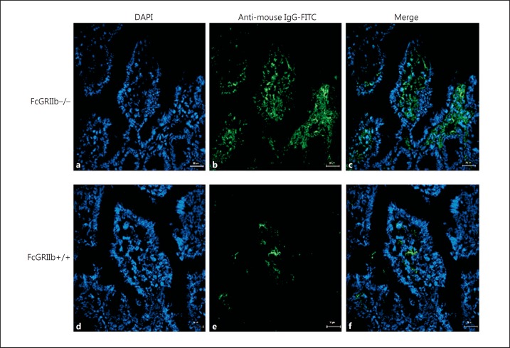
Fig. 3.
Representative immunofluorescence images from the jejunums of 40-week-old FcGRIIb−/− mice (a–c) versus wild-type controls (FcGRIIb+/+) (d–f). ×600. DAPI (blue) and goat anti-mouse IgG with FITC (green) were used for the identification of the nucleus and Fc portion of the immune complex deposition, respectively. The merged figures demonstrated that immune complex depositions were mostly found at the lamina propria of FcGRIIb−/− mice (c) when compared with wild-type mice (f).

