Fig. 4.
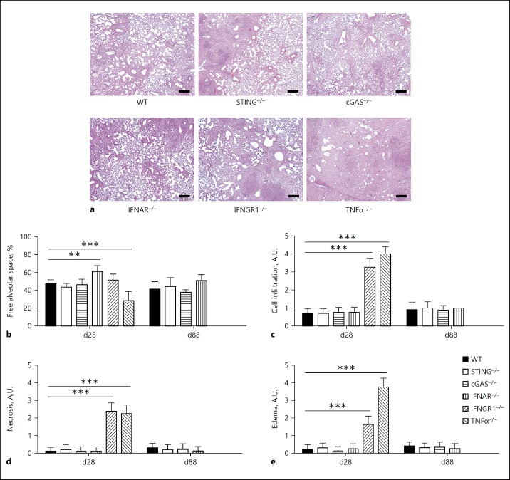
Absence of STING does not influence lung pathology during infection by Mtb. Lungs from WT, STING–/–, cGAS–/–, IFNAR–/–, IFNGR1–/–, and TNFα–/– mice infected with Mtb H37Rv (as in Fig. 3) were collected at 28 or 88 days postinfection (d28 or d88), fixed and embedded in paraffin. a Representative pictures of stained slides from lungs after 28 days of infection. H&E. Scale bar, 100 µm. b The free alveolar space was evaluated using ImageJ software and expressed in relation to the total area analyzed. Vascular vessels, bronchi, and bronchioles were excluded from the analysis. Cell infiltration (c), necrosis (d), and edema (e) were also scored from each slide. The results are expressed as arbitrary units (A.U.), ranging from 0 to 5. The IFNGR1–/– and TNFα–/– mice did not survive for 88 days. Data are shown as mean ± SD of n = 5 mice per group and the results presented are from 1 experiment representative of 3 independent experiments performed. Statistically significant difference in relation to WT: * p < 0.05; ** p < 0.01; *** p < 0.001 (two-way ANOVA followed by the Bonferroni post hoc test).

