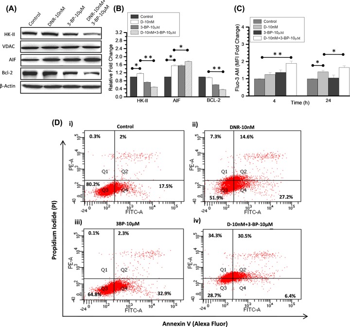
Figure 5. Mitochondrial HK-II dissociation accompanied by increased extracellular calcium and apoptosis in combined treatment.
(A) Immunoblotting was performed (at 4 h) to examine the treatment-induced changes in HK-II expression level and its correlation with initial apoptotic signals in K-562 cells. HK-II protein expression presented here in the mitochondrial enriched fraction of control, DNR (10 nM), 3-BP (10 μM) and their CT compared with loading control VDAC. Apoptosis inducing factor (AIF) and anti-apoptotic protein (Bcl-2) were checked in the whole cell lysate and compared with respective β-actin. (B) Immunoblotting densitometry was performed (using ImageJ software) in the indicated proteins and graph presented as relative fold change compared with control. (C) Intracellular calcium measurement was carried out using Fluo-3AM at indicated time points after the DNR and 3-BP treatment in combination and alone. Graph presented here as relative fold change in the mean fluorescence intensity (MFI) with respective control. Data are expressed as mean ± SD (n=3), statistical significance (*P<0.05; **P<0.01 vs. untreated control cells). (D) Flow cytometric analysis using Annexin V (Alexa fluor)/PI in the combined (DNR and 3-BP) and TA in K-562 cells at 24 h showing retention of different apoptotic phase events (%) presented as quadrants (Q1–Q4).

