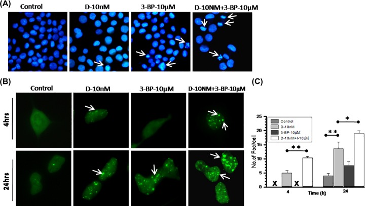
Figure 6. DNR-induced DNA damage significantly enhanced with 3-BP combined treatment.
(A) Nuclear fragmentation assay was performed to visualize the treatment induced alteration in nuclear morphology in K-562 cells. At 24 h, post treatment of DNR and 3-BP alone and in combination cells were fixed and stained with DAPI. Photomicrograph showing variation in nuclear size, condensed chromatin, and fragmented nuclei compared among treatment groups. Images were captured under 10× (objective) × 10× (eyepiece) magnification. (B) DNA damage analysis was carried out at indicated time points post-treatment of DNR and 3-BP alone and combination in HEK-293 cells transfected with DNA damage marker 53-BP1-GFP. In photomicrograph, arrow indicates foci formation in damaged cells. (C) From image analysis number of foci counted and graph presented as number of foci per cell compared among treatment groups. All the images were captured under 20× (objective) × 10× (eyepiece) magnification. Quantitative data are expressed as mean ± SD (n=3) and statistical significance *P<0.05; **P<0.01 vs. untreated control cells.

