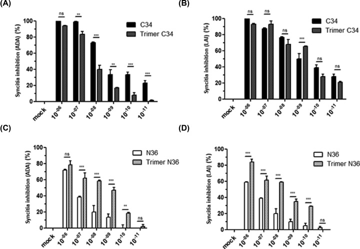
Figure 3. Characterization of the anti-fusion activity of C3 and N36 peptides.
HeLa cells expressing envelope glycoproteins from either HIV-1 ADA (R5-tropic virus) (A,C) or HIV-1 LAI (X4-tropic virus) (B,D) were incubated with HeLa cells expressing human CD4 receptor and CXCR4 or CCR5 HIV-1 co-receptors in the presence of escalating concentrations (10−12–10−6 M) of N36 or C34 peptides either in monomeric or trimeric conformation. After 20 h of incubation, syncytia formation was quantified by optical microscopy. The percentage of inhibition of syncytia formation was calculated. ‘mock’ corresponds to the syncytia formation obtained in the absence of peptide inhibitors but including the same medium as that used for the solubilization of the peptide inhibitor tested. Experiments were performed in triplicate and repeated three times. A representative experiment is shown as mean ± standard deviation. **, P<0.01; ***, P<0.0001

