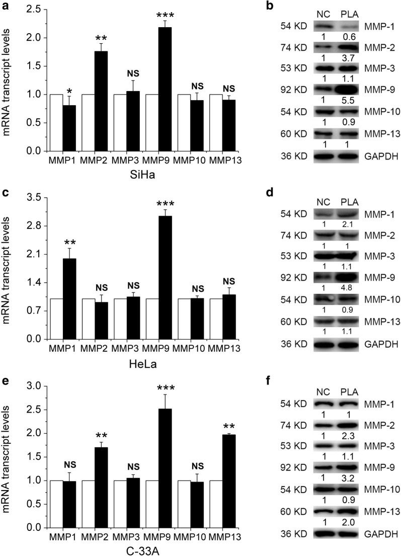
Fig. 2.
Effects of PLA on the expression of migration- and invasion-related proteins in cervical cancer cells. MMP expression was detected by real-time PCR analysis in SiHa (a), HeLa (c), and C-33A (e) cells treated with and without PLA (10 mM) for 24 h. Data are presented as the means ± SDs of three independent experiments. *P < 0.05, **P < 0.01, and ***P < 0.001 compared with the control. After treatment with PLA for 24 h, the expression of MMP-1, MMP-2, MMP-3, MMP-9, MMP-10 and MMP-13 in SiHa (b), HeLa (d), and C-33A (f) cells was detected by Western blot analysis. GAPDH expression was evaluated as a loading control. One representative of three different experiments, for each of the analyses performed, is shown

