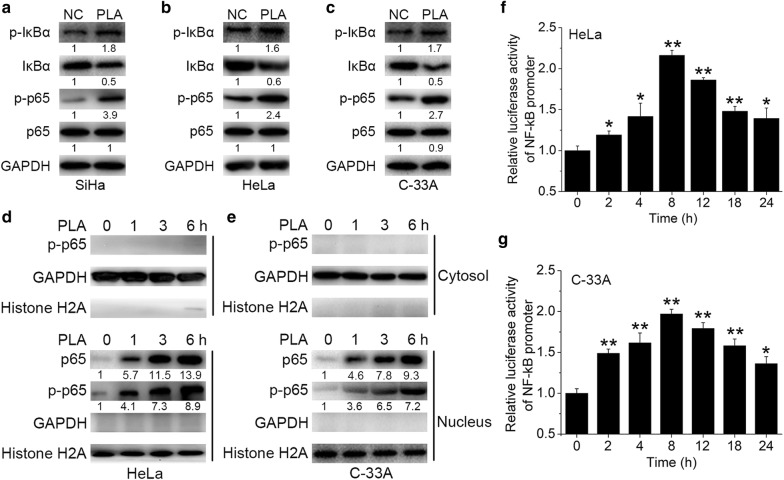
Fig. 4.
Effects of PLA on NF-κB activation in cervical cancer cells. a‒c SiHa (a), HeLa (b), and C-33A (c) cells were treated with 10 mM PLA for 30 min, and the activation of IκBα and p65 was then examined by Western blot analysis. GAPDH expression was evaluated as a loading control. One representative of three different experiments, for each of the analyses performed, is shown. d, e) After treatment with 10 mM PLA for the indicated times, the nuclear and cytoplasmic cellular fractions of HeLa (d) and C-33A (e) cells were isolated by differential lysis. The levels of p-p65 in the nuclear and cytoplasmic cellular fractions were detected by Western blot analysis. GAPDH and Histone H2A were used as loading controls. One representative of three different experiments, for each of the analyses performed, is shown. f, g HeLa (f) and C-33A (g) cells were cotransfected with pNF-κB-Luc and pRL-TK. After transfection for 6 h, cells were treated with 10 mM PLA for the indicated times. NF-κB reporter activity was calculated as the ratio of the activities of pNF-κB-Luc and pRL-TK. Data are presented as the means ± SDs of three independent experiments. *P < 0.05 and **P < 0.01 compared with the control

