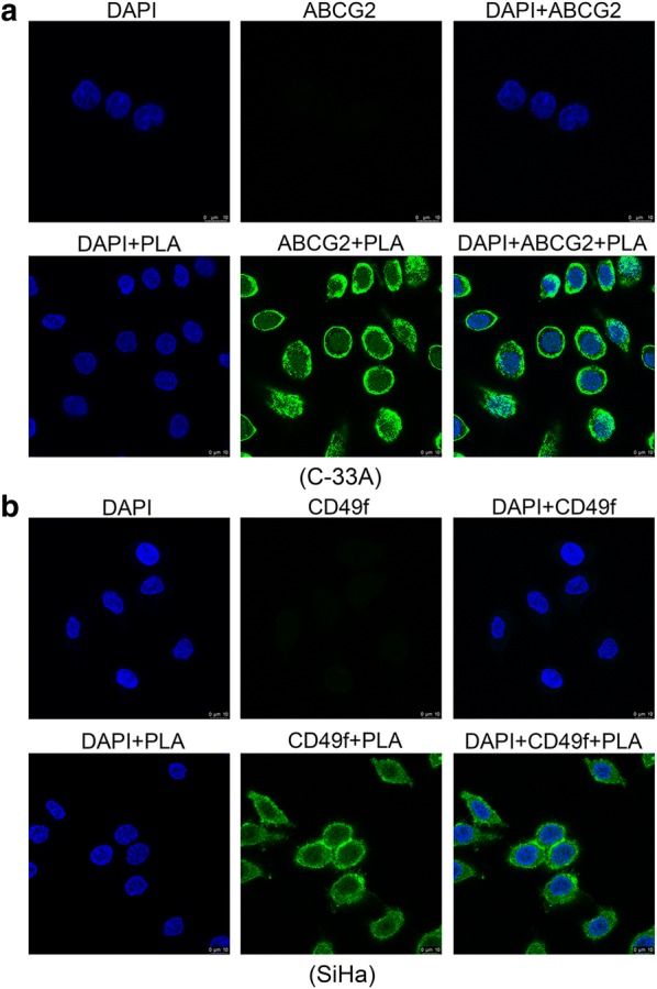
Fig. 6.

Fluorescence microscopy analysis of the labeled cervical cancer stemness markers. C-33A (a) and SiHa (b) cells were treated with or without PLA (10 mM) for 12 h, and then, ABCG2 and CD49f proteins were examined by immunostaining. DAPI (4′,6-diamidino-2-phenylindole) staining was applied as a nuclear marker. Scale bar, 10 μm
