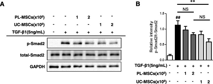
Fig. 7.
UC-MSCs inhibit TGF-β1-induced phosphorylation of Smad2. HIMFs were treated with TGF-β1 (5 ng/mL) and co-cultured with or without UC/PL-MSCs at 1 or 2 × 10⁵ cells/insert for 48 h. a Representative Western blots show the protein expression of phosphorylated Smad2 (p-Smad2) with total Smad2 and GAPDH as a loading control. b Quantitation of p-Smad2 from Western blot analyses (n = 4). Data are expressed as the means ± SEM. ##P < 0.01 versus the untreated control; **P < 0.01 versus the TGF-β1 treatment only. NS, not significant

