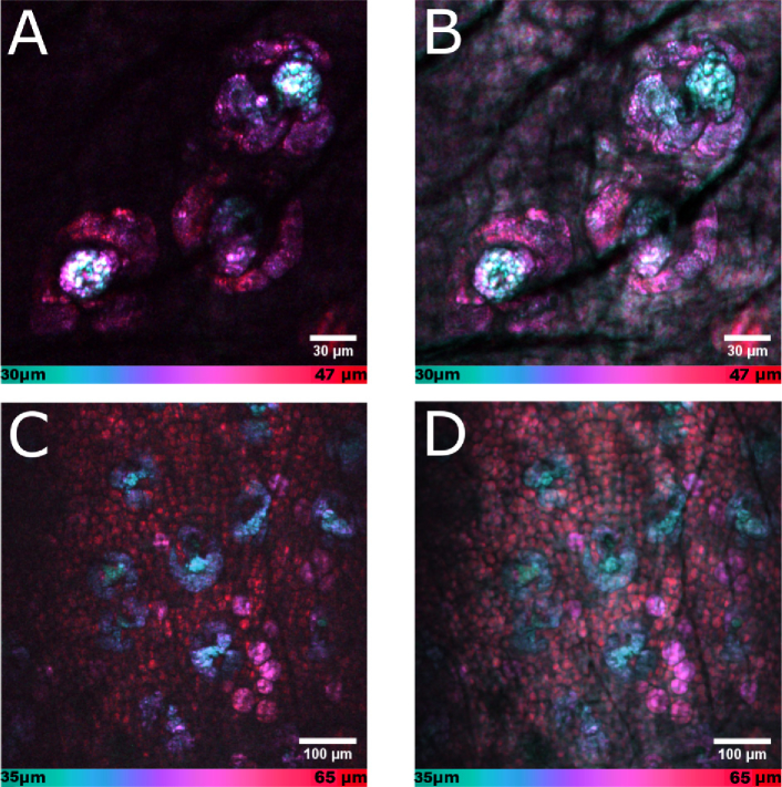
Fig. 6.
Color-depth projections of 3D volumes of fresh ex vivo mouse ear tissue imaged with lipid contrast ( = 2845 ) CARS. Reference point of the depth color code bars at the bottom of each image is the skin surface at 0 m. Volumes were consequently obtained with the fiber-based light source (a, c) and a reference light source (b, d). (a, b) Lipid-rich sebaceous glands. (c, d) Dermal structures from the sebaceous glands to adipocytes, and subcutaneous fat. The spectrally broadband excitation with the femtosecond reference source resulted in a higher overall brightness but lower image contrast due to the stronger non-resonant signal contribution in (b) and (d).

