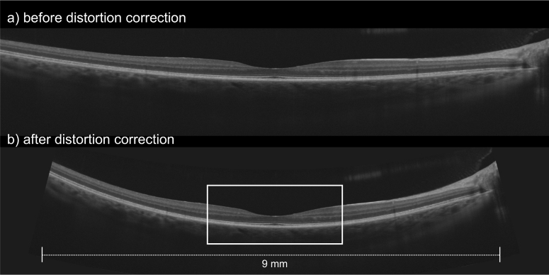
Fig. 1.
Comparison of an OCT line scan (a) before and (b) after distortion correction [17]. Both scans are of the same original scan length of approximately 9 mm (depending on the magnification factor given by the axial length). The white box in part (b) indicates the region of interest for the analysis of foveal pit morphology.

