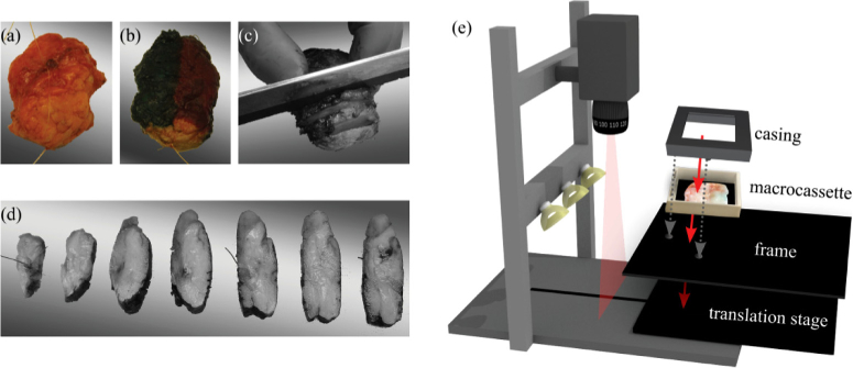
Fig. 2.
Data acquisition of breast tissue slices. The breast specimen before (a) and after (b) inking with histopathologic ink. The specimen was sliced (c, d), and one slice was selected and placed in a macrocassette for optical measurements. The tissue slice was imaged with both HS imaging systems (e). To allow for a reproducible location for each measurement and an accurate registration between both cameras, the macrocassette with the tissue was fixed with a casing on a frame that fitted the translational stage of both systems.

