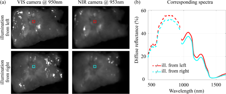
Fig. 6.
Intensity differences between spectra. (a) Hyperspectral images obtained with the VIS and NIR camera. In the top and bottom row, the tissue was illuminated from the left and the right, respectively. The colored squares in the HS images are located at the same position in the tissue and correspond to the diffuse reflectance spectra in (b). Spectra obtained with different cameras (VIS and NIR) did not connect due to differences in the measurement setup of both cameras in combination with the rough surface of the tissue slices. This might cause a spectral variability that can be observed as a baseline shift of the spectra. Even when diffuse reflectance spectra were taken with the same camera, at the same location in a tissue slice, their intensity varied when the tissue was illuminated from a different point of view.

