
Fig. 3.
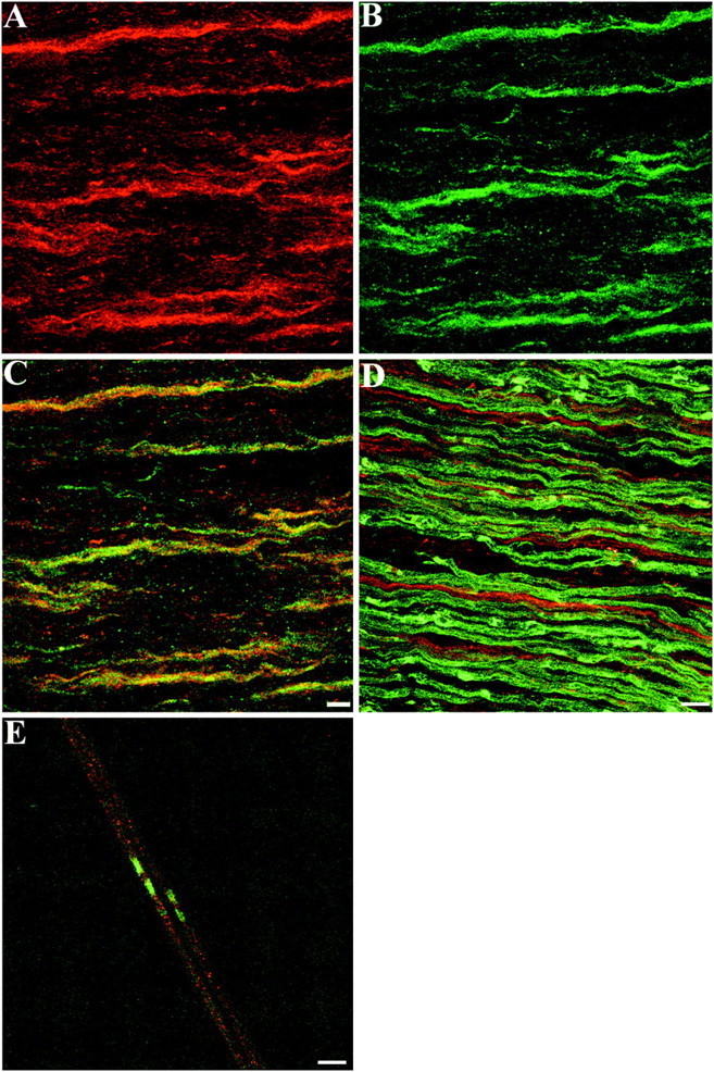
Distribution of TAG-1 in Schwann cells of postnatal sciatic nerve. A, On P6 sciatic nerve sections, TAG-1 is expressed by ensheathing Schwann cells detected by L1 labeling (B, green). Cis the combined image of A and B that shows that both channels overlap (yellow).D, On P6 sciatic nerve sections, myelinating Schwann cells detected by MAG labeling (green) are negative for TAG-1 (red), which appears in ensheathing Schwann cells only. E, At P6 myelinated fibers, TAG-1 (red) does not appear to be clustered, whereas in the paranodal region paranodin/Caspr is already detected (green). A–C, Eleven images, 0.6 μm apart; D, nine images, 0.9 μm apart;E, seven images, 0.6 μm apart. Scale bar, 10 μm.