
Fig. 6.
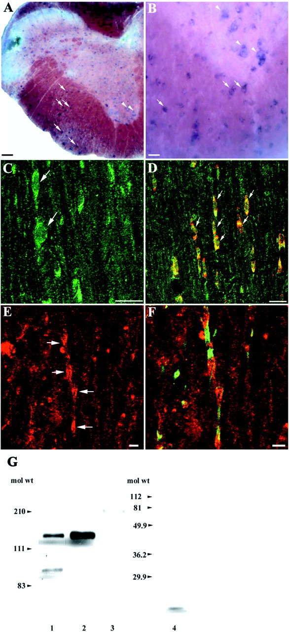
TAG-1 is expressed by oligodendrocytes and accumulates at the juxtaparanodal region. A, In adult spinal cord sections, TAG-1 mRNA is expressed in small cells of the white matter (arrows, higher magnification inB). In the ventral horn TAG-1 is also detected in large, spindle-shaped neurons (arrowheads, higher magnification in B). C, In the white matter of adult spinal cord sections, TAG-1 protein is present in the soma of round to oval cells (green, arrows). D, In these cells, TAG-1 (green) colocalizes with APC (red), a marker for oligodendrocytes, as both channels overlap (yellow, arrows). E, TAG-1 also appears in clusters in white matter (red, arrows).F, Sequential labeling for paranodin/Caspr (green) shows that TAG-1 (red) has a discrete distribution and is concentrated in the juxtaparanode of myelinated fibers. G, TAG-1 protein is detected in myelin-rich membrane fractions isolated from adult spinal cord as a 135 kDa band and a 90 kDa adult isoform (lane 1). Whole protein extract from postnatal cerebellum was loaded in lane 2. Low levels of L1 protein are detected in the above myelin fractions (180 kDa band, lane 3). In these fractions, MBP (18.5 kDa band, lane 4) is abundantly expressed because it is detected in of the amount loaded inlanes 1 and 3. C, Ten images, 0.4 μm apart; D, 5 images, 0.4 μm apart; E,F, 11 images, 0.9 μm apart. Scale bars: A, C, D, 20 μm; B, E, F, 5 μm.