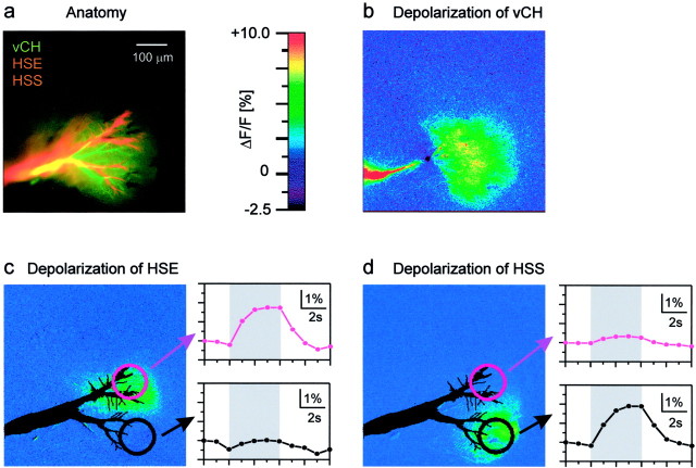
Fig. 4.
Dendrotopic calcium accumulation in a vCH cell.a, Anatomy of a vCH cell filled with Calcium Green and an HSE and an HSS cell both filled with the red fluorescent dye Alexa.b–d, False-color images of the fluorescence change in the vCH cell. In c and d, the outlines of the HSE and HSS cells are superimposed in black. Also shown are the time courses of the relative change of fluorescence within the areas outlined in c andd during injection of depolarizing current into the HSE (c) and into the HSS (d) cell (lens, Zeiss UC 20×, 0.57 NA).

