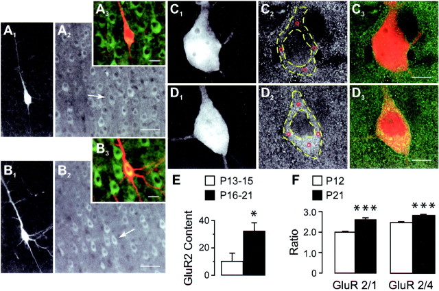
Fig. 2.
GluR2 expression in pyramidal neurons at different ages. A, B, Dual-channel images of recorded biocytin-filled layer 5 pyramidal neurons from P14 (A1–A3) and P18 (B1–B3) animals immunostained with the GluR2 antibody (A1,B1, biocytin-Texas Red;A2, B2, GluR2-FITC;A3, B3, color overlap). Note that pyramidal neurons, including biocytin-filled cells (A2, B2,arrows), in the older age group show higher levels of GluR2 immunofluorescence compared with younger under similar conditions of illumination. Scale bars: A1,A2, B1,B2, 50 μm;A3, B3, 20 μm.C, D, High-magnification images of young (C1–C3) and old (D1–D3) biocytin-filled neurons showing that although the intracellular level of GluR2-FITC fluorescence in the young animal (C2) is comparable with adjacent neuropil and with the intranuclear staining, it is markedly more intense in the corresponding regions of the older neuron (C2 vs D2). Note the absence of cross-talk between Texas Red (C1,D1) and FITC (C2,D2) channels. Immunofluorescence was measured at four locations (C2 andD2, red circles) within the perikaryon (outlined with dashed yellow lines), and the average of these values was used to represent cell brightness (see Materials and Methods). Scale bars, 10 μm. E, GluR2 levels in P13-P15 (n = 6) and P16-P21 (n = 9) biocytin-filled pyramidal neurons based on perikaryon immunofluorescence measurements compared with background as outlined in C, D. F, Changes in GluR2 immunoreactivity relative to coexpressed GluR1 (or 4) measured separately in a population of layer 5 pyramidal cells from corresponding cortical regions in intact brain sections of animals at the indicated ages (P12, n = 142 cells, 11 sections; P21, n = 83 cells, 10 sections). *p < 0.05; ***p < 0.0001.

