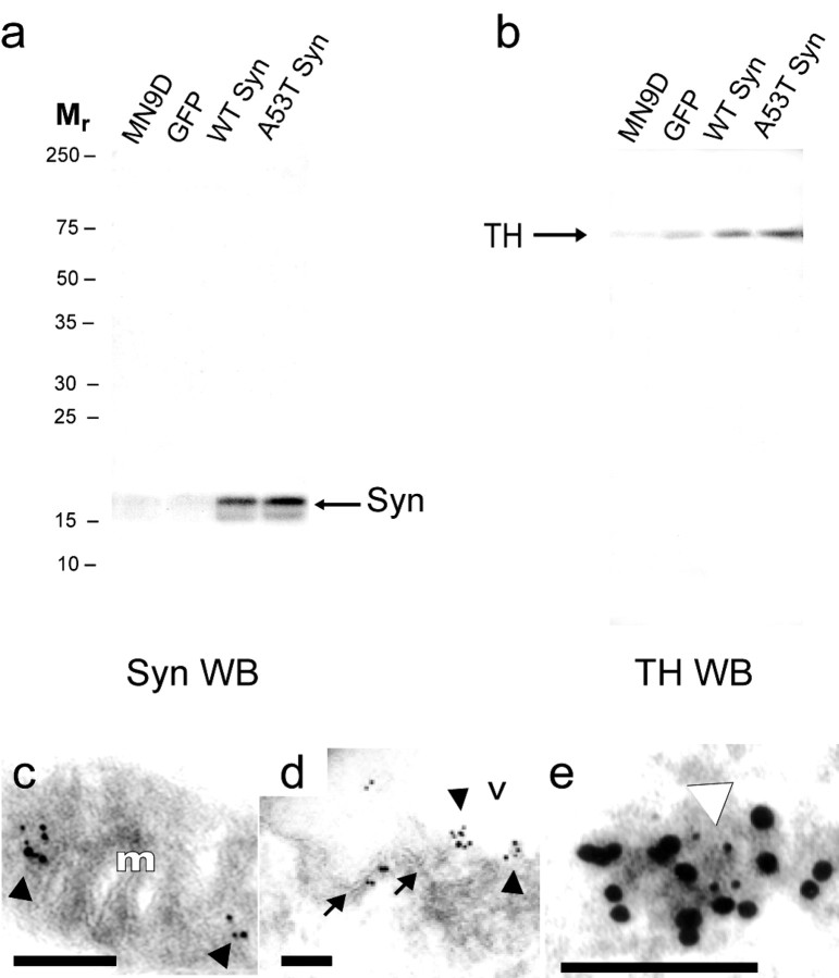
Fig. 4.
Interaction of α-synuclein with TH in MN9D cells. Western blots (WB) from a representative co-IP experiment reacted with the anti-α-synuclein antibody (a) or with the anti-TH antibody (b). Immunoprecipitation of α-synuclein from cell extracts (a) resulted in co-IP of TH (b) from parental MN9D (MN9D), GFP-transfected MN9D (GFP), wild-type (WT) α-synuclein, and A53T α-synuclein (A53T Syn) cells. Immunoelectron microscopy reveals colocalization of TH with α-synuclein that is apparent on mitochondria (c) and vesicles (d,e) in MN9D cells. In c andd, the larger (10 nm) gold particles label α-synuclein and the smaller (5 nm) particles label TH colocalized on the surface of a mitochondrion (c) and at the edge of a vesicular structure (d) in an MN9D cell stably transfected with A53T α-synuclein. In e the large (10 nm) gold particles label TH and the small (5 nm) particles label α-synuclein in the cytoplasm of an MN9D cell stably transfected with wild-type α-synuclein. Arrowheads inc–e point to colocalized small and large gold particles. Arrows in d point to the lipid bilayer of a vesicle decorated with large and small gold particles. m, Mitochondrion; v, vesicle. Scale bars, 100 nm.

