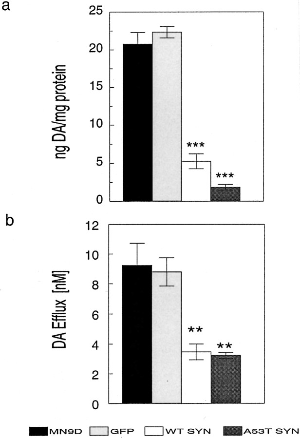
Fig. 5.
Effect of α-synuclein overexpression on cellular DA and DA efflux from MN9D cells. Supernatants obtained from cell lysates of parental MN9D, GFP, wild-type α-synuclein (WT SYN), and A53T mutant α-synuclein cells were assayed for DA using HPLC with electrochemical detection (a). Although A53T cells had even lower cellular DA levels than wild-type α-synuclein cells in a, the difference was not statistically significant using ANOVA with Tukey-Kramer post hoc analyses (p > 0.05). To measure DA efflux from the various MN9D lines, cells were washed in ACSF and incubated in fresh ACSF for 15 min at 37°C before collection (b). Data, normalized for total protein, represent the mean ± SEM of triplicate samples from two to six independent experiments.Black bar, MN9D; light gray bar, GFP;white bar, wild-type α-synuclein; dark gray bar, A53T α-synuclein. **p < 0.01; ***p < 0.001.

