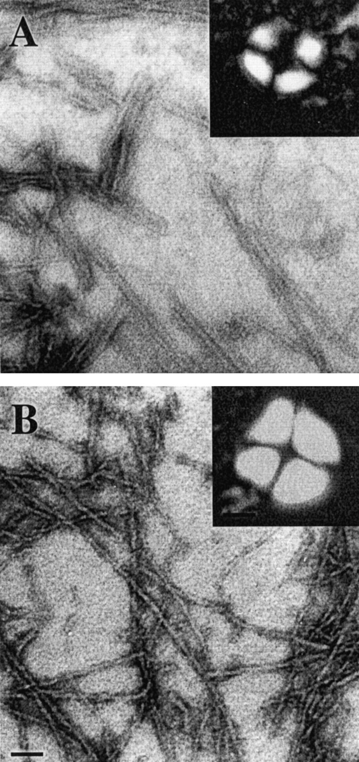
Fig. 2.

Electronic micrographs of negatively stained AβGlu22→Gln fibrils (A) and AβGlu22→Gln–AChE complexes (B). Scale bar, 0.15 μm. Insets are pictures of Maltese crosses obtained for both AβGlu22→Gln fibrils (A) and AβGlu22→Gln–AChE complexes (B) by staining with Congo red. Pictures were taken with a Nikon Optiphot microscope configured for polarized light. Scale bar, 15 μm.
