Fig. 5.

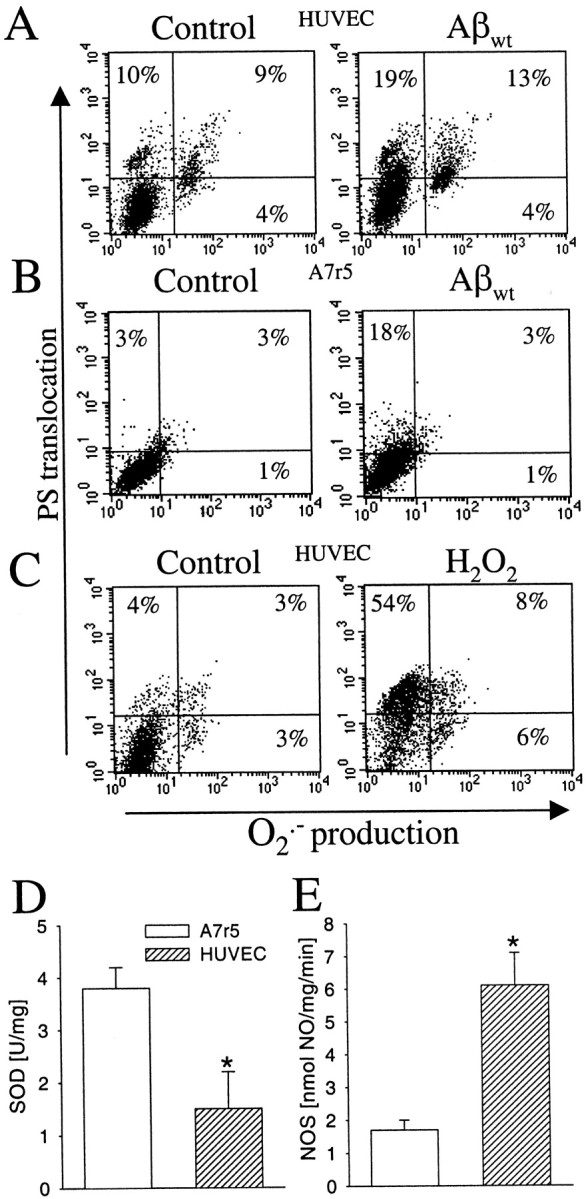
Oxidative stress and Aβ-mediated vascular cytotoxicity. Phosphatydylserine translocation to the outer membrane and O2·− production on HUVEC cells (A) and A7r5 cells (B) exposed to 10 μm Aβwt fibrils and HUVEC cells (C) with 50 μmH2O2 at 48 hr. Percentages indicate cells positive for annexin-V binding (top quadrants) and O2·− production (right quadrants) in regard to the controls (bottom left quadrant). Activity of SOD (D) and NOS (E) in A7r5 and HUVEC cells. Results are expressed in relation to milligrams of protein. Data are mean ± SEM (error bars) values of seven to eight cell samples analyzed in triplicate. *p < 0.05 by nonpaired Student'st test versus the respective results on A7r5 cells.
