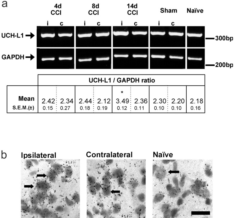
Fig. 3.

Determination of levels of mRNA for ubiquitin C-terminal hydrolase-L1 (UCH-L1). a, Relative abundance of UCH-L1 mRNA from spinal cord of CCI-treated rats as assessed by semiquantitative RT-PCR normalized to the signal obtained for the cellular housekeeping enzyme GAPDH. Densitometric analysis indicated that the relative UCH-L1 expression was similar in all conditions except in tissue ipsilateral to CCI at 14 d, where expression was significantly greater than that in contralateral samples (*p < 0.05 by paired Student's ttest; n = 3). Further analysis of the changes at 14 d after CCI was made by in situ methods. Seeb and Table 2 for quantification data. b,In situ hybridization histochemistry to show UCH-L1 mRNA expression in lamina I of the rat medial dorsal horn. High-power, light-field photomicrographs showing that expression of mRNA for UCH-L1 was higher ipsilateral to CCI after 14 d, compared with contralateral tissue and naive dorsal horn. Scale bar, 100 μm.
