Fig. 4.
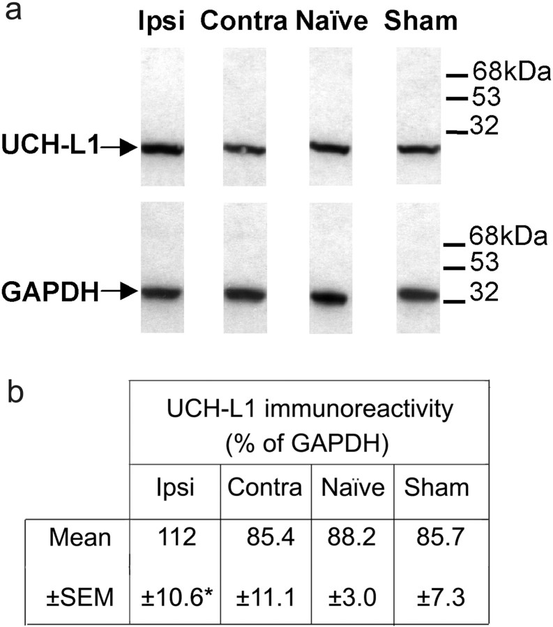
Western blot analysis of the expression of UCH-L1 protein. Western blots of spinal cord samples from neuropathic, naive, and sham-operated rats. a shows an increase in UCH-L1 protein expression ipsilateral (Ipsi) to CCI in neuropathic animals. No change from naive samples was observed in the sham-operated or contralateral side (Contra) of CCI samples. b represents UCH-L1 expression as a percentage of GAPDH expression in terms of relative gray scale values after quantitative densitometry of ECL films. Data are presented as mean ± SEM (n = 3; *p < 0.05, from control contralateral values; Student's paired ttest).

