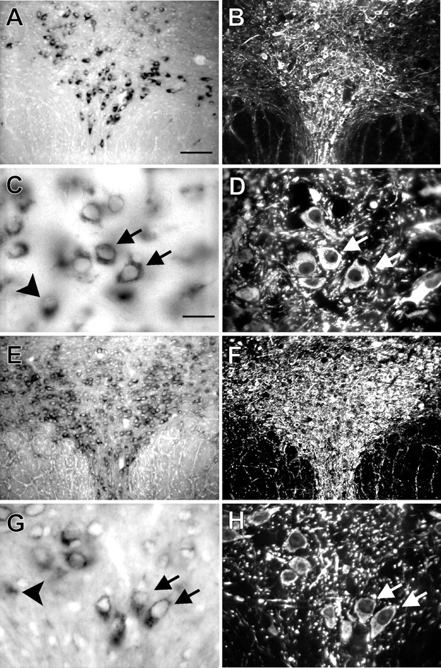
Fig. 3.

TASK-1 and TASK-3 transcripts are expressed in TPH-IR neurons of the dorsal raphe. In situhybridization with digoxigenin-labeled RNA probes complementary to TASK-1 and TASK-3 was combined with immunohistochemistry for TPH on coronal sections of rat midbrain at the level of RDo. Low-powered bright-field images show labeling for TASK-1 (A) and TASK-3 (E); fluorescence photomicrographs of the same sections reveal immunostaining for TPH (B,F). Note the strong correspondence in localization of TASK-expressing and TPH-immunoreactive (i.e., serotonergic) cells. High-magnification bright-field images show individual TASK-1-labeled neurons (C) and TASK-3-labeled neurons (G); immunofluorescence photomicrographs identify TPH-IR neurons in the same sections (D, H). It is clear that most serotonergic neurons express TASK channels (arrows); some TASK-expressing neurons that are not noticeably TPH-IR were also apparent (see arrowheads in C andG). Scale bars: A, 100 μm;C, 25 μm. Sections in A–D were taken from a slightly different rostrocaudal level than those inE–H.
