
Fig. 5.
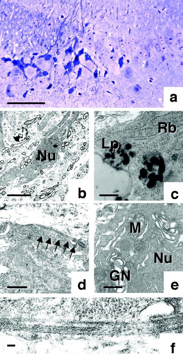
Ultrastructural analysis of irregularly shaped neurons. a, Light photomicrograph of a toluidine blue-stained ultrathin section from the hippocampus of a Tg mouse, showing irregularly shaped neurons. b, Low-power electron photomicrograph of hippocampal neurons from a Tg214 mouse, showing an example in the center of dark cell degeneration.c, Examination of this electron-dense cell at a higher magnification clearly shows the accumulation of ribosomes (Rb) and lipofuscin (Lp).d, The presence of bundles of straight tubules in the cytoplasm of this neuron is also evident at higher magnifications. Further examination of these tubules at a higher magnification (f) confirms that the diameter of these bundles is ∼15 nm. e, Higher magnification reveals the swelling of Golgi network (GN) and the ruffling of nuclear (Nu) membrane. Scale bars: a, 60 μm; b, 5 μm; c, 350 nm;d, 1 μm; e, 160 nm; f, 100 nm.