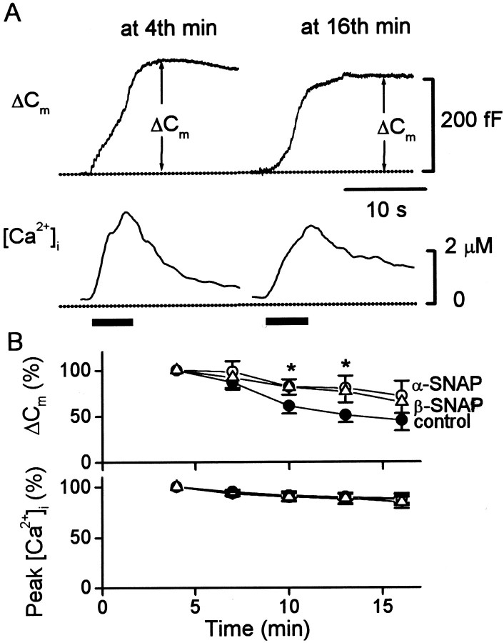
Fig. 9.
Effects of α- or β-SNAP on amplitude and time-dependent rundown of the exocytic response. A, Amplitude of the exocytic response (measured as increase in membrane capacitance, Cm) triggered by a train of 15 depolarizations (indicated by a bar below the traces) delivered at the 4th and 16th min after establishment of whole cell. The cell was recorded with 500 nm β-SNAP in the pipette solution. The train of depolarization delivered at 4 min of whole cell increased the membrane capacitance by 233 fF, corresponding to a 5.8% increase in cell membrane capacitance (initial cell capacitance = 4 pF). At 16 min of whole cell, the train of depolarization triggered a ∼25% smaller increase inCm, and the rise in [Ca2+]i was also slightly smaller.B, Plot of the exocytic response and peak [Ca2+]i triggered by trains of depolarization delivered at the 7th, 10th, 13th, and 16th minute of whole cell dialysis. The values of the exocytic response and peak [Ca2+]i were normalized to the values of the individual cell obtained at the fourth minute, which had no statistical difference between control cells (●) and cells dialyzed with 500 nm α-SNAP (○) or β-SNAP (▵). Theasterisks indicate that the data for α- or β-SNAP are statistically different from control cells at the same duration of dialysis. Note that exogenous α- or β-SNAP (500 nm) significantly reduced the time-dependent rundown in the exocytic response between the 10th and 13th minute but had no significant effect on the smaller rundown in peak [Ca2+]i. However, there is no significant difference between the data for α- and β-SNAP.

