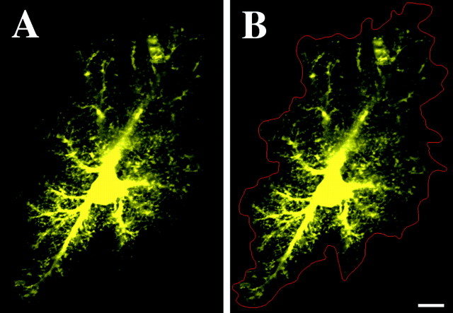
Fig. 1.
Protoplasmic astrocyte of CA1 stratum radiatum iontophoretically filled with the fluorescent dye Alexa 488.A, Optical slice reveals the dense spongiform processes of these astrocytes. A clear but complex boundary is evident in the extent of these processes. B, Via manual segmentation (red), it was possible to delineate the extent of and calculate the neuropilar volume occupied by each astrocyte. Scale bar, 10 μm.

