
Fig. 8.
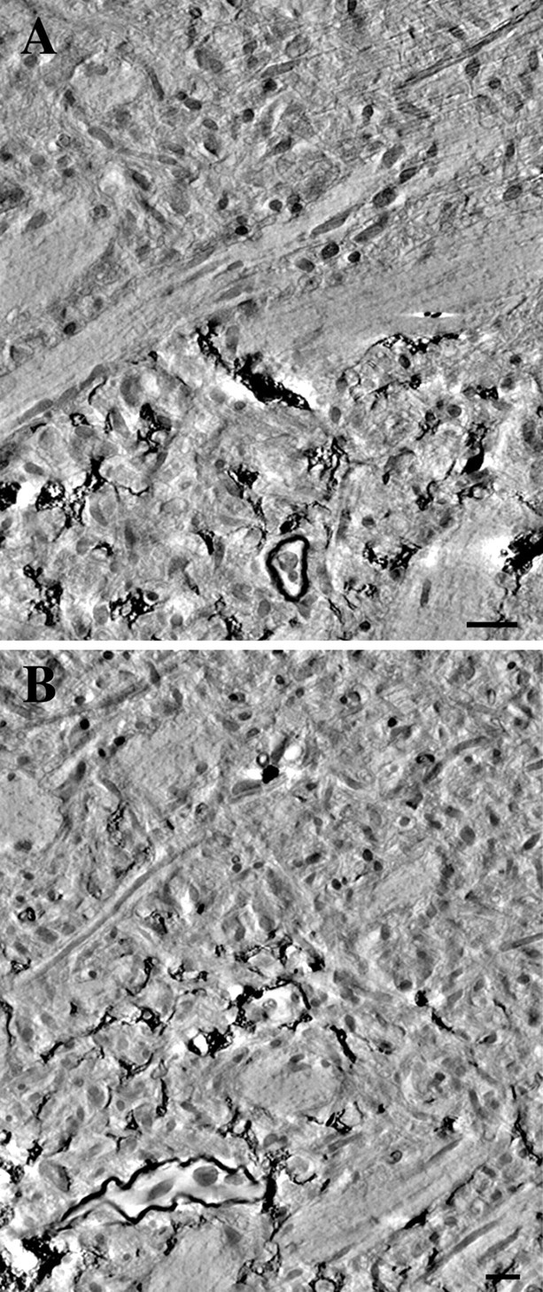
Electron microscopic examination of the boundary regions of a photooxidized astrocyte. A,B, Tomographic reconstructions of astrocyte boundary regions were generated using a 0.5-μm-thick section through a photoconverted astrocyte. Computational slices through the resulting volumes demonstrate an abrupt decrease in the density of fine astrocytic processes at the boundary to extent of the astrocyte. Scale bars, 1 μm.